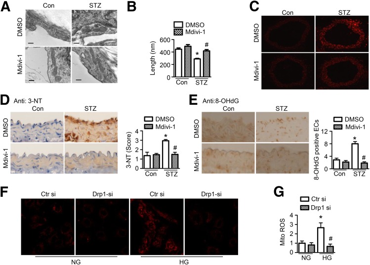
Figure 6.
Inhibition of mitochondrial fission attenuates oxidative stress in diabetic aorta. A–F: STZ-induced diabetic mice were treated with mdivi-1 (1.2 mg/kg/d) or vehicle (DMSO) using an osmotic pump for 14 days. A: Representative transmission electron micrographs of mitochondria in the aortic endothelium. Scale bars = 300 nm. B: Quantification of average mitochondrial length. n = 6 mice, at least 50 mitochondria per mice were analyzed. *P < 0.05 vs. control mice; #P < 0.05 vs. DMSO. C: Frozen sections of aortas were incubated with 5 μmol/L DHE for 30 min. Images were obtained at 518 nm (excitation) and 605 nm (emission). n = 5. D: Representative images of immunohistochemical staining and quantification of positive staining for 3-NT in thoracic aortic sections. n = 5. *P < 0.05 vs. control; #P < 0.05 vs. vehicle. E: Representative images of immunohistochemical staining for 8-OHdG in aortas. Quantification of 8-OHdG–positive endothelial cells (ECs). n = 5 mice. *P < 0.05 vs. control mice; #P < 0.05 vs. DMSO. F and G: HUVECs were transfected with Drp1 siRNA (si) or control siRNA for 24 h, after which they were treated with high glucose (HG) for 24 h. Mitochondrial ROS production was measured by incubating HUVECs with 2 μmol/L MitoSOX for 30 min. F: Representative fluorescence images are shown. G: Quantification of fluorescence intensity for mitochondrial ROS levels in HUVECs. n = 6. *P < 0.05 vs. normal glucose (NG); #P < 0.05 vs. control siRNA.

