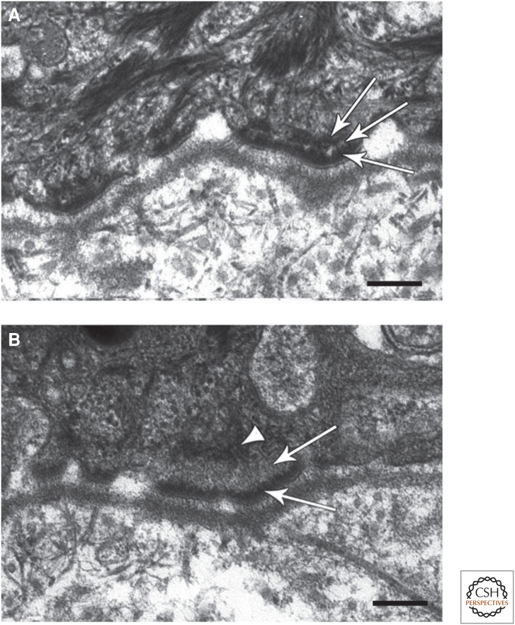
Figure 5.
A role for BPAG1 in mediating anchorage of intermediate filaments (IFs) at hemidesmosomes (HDs). (A) Electron micrograph of a section of typical human skin at the region of interaction between keratinocytes and the basement membrane zone (BMZ). Three HDs are shown, each possessing a tripartite plaque (arrows). Note that IFs show extensive interaction with the inner HD plaque. (B) Electron micrograph of a comparable region of the skin of a patient showing loss of expression of bullous pemphigoid antigen 1 (BPAG1). The arrowhead indicates that the HD plaque lacks an obvious inner layer, and the arrows mark the keratin IF interaction with the HD that is also abnormal. Scale bar, 100 nm. (Images kindly provided by John McGrath, King’s College, London, and James McMillan, St Thomas’ Hospital, London). (A,B Adapted from Groves et al. 2010, with permission from Macmillan Publishers Ltd.)

