Figure 6.
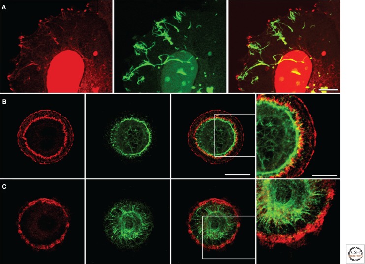
Interactions between focal adhesions (FAs) and the cytoskeleton. (A) Human breast adenocarcinoma MCF7 cells induced to express vimentin tagged with green fluorescent protein and then fixed and stained with antibodies against β3 integrin (red). The cells, when made to express vimentin, initiate filament assembly at FAs in the periphery of the cells. The prominent nuclear stain is an artifact. Scale bar, 10 µm. Primary lung alveolar cells were treated with (B) control adenovirus or with (C) adenovirus encoding short hairpin RNA targeting plectin expression, plated on a micropatterned surface comprising a series of matrix-coated circles, as detailed elsewhere (Eisenberg et al. 2013), and then costained for the FA protein talin (red) and the intermediate filament protein keratin (green). Bar, 20 µm (panel 3). Note how knockdown of plectin not only inhibits the assembly of the perinuclear FAs but appears to “release” the keratin cytoskeleton, which then makes contact with FAs at the cell periphery. The boxed areas in the third panels in B and C are shown at higher power in the fourth panel (scale bar, 10 µm).

