
Figure 6.
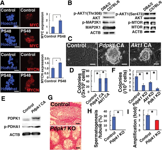
Crucial role of the PDPK1–AKT–PDK1 pathway in SSC self-renewal. (A) Immunostaining of B6-GS cells for MYC (n = 16 for PS48; n = 14 for control) and MYCN (n = 23 for PS48; n = 26 for control) expression. Testis cells from 8-d-old pups were cultured overnight on gelatin-coated plates and transferred on MEFs for 4 d before staining with the indicated antibodies. Signal intensity was measured by dividing total signals by colony area. (B) Western blot of 8-d-old DBA/2 and B6 germ cells after overnight culture with GDNF and FGF2. (C) Appearance of B6-GS cells established by transfection of constitutively active Pdpk1 (Pdkp1 CA) or Akt1 (Akt1 CA). (D) Quantification of in vitro colony formation by B6 germ cells after transfection of Pdkp1 CA or Akt1 CA. n = 4. Germ cell clumps were enumerated 37 d after transfection of 3 × 10⁵ B6 germ cells. (E) Western blot of PDHA1 after Pdpk1 CA transfection in B6-GS cells 3 d after transfection. (F) Colony counts in recipient mouse testes that underwent transplantation of testis cells transfected with lentivirus vectors expressing shRNA against Pdpk1, Akt, or Pdk1. n = 18 for control; n = 12 for Pdpk1; n = 13 for Akt; n = 12 for Pdk1. (G) Histological appearance of recipient testes transplanted with Pdpk1f/f testis cells after exposure to AxCANCre. (H) Quantification of seminiferous tubules with spermatogenesis. At least 354 tubules in four testes were counted. (I) Proliferation of Pdpk1f/f GS cells after AxCANCre treatment. Cells were infected with AxCANCre and passaged at 5 d after infection. Cell recovery was determined 7 d after the passage. n = 3. Bars: A,C, 20 µm; G, 50 µm. The asterisk indicates a significant difference.