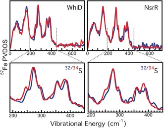
Figure 2.

NRVS spectra of [4Fe‐4S] WhiD and NsrR. Top: 57Fe PVDOS for: Left (blue) unlabeled (32S) WhiD and (red) 34S‐labeled WhiD; right (blue) unlabeled (32S) NsrR and (red) 34S‐labeled NsrR. Bottom: Magnified view of the 200–450 cm−1 regions of the 32/34S WhiD (left) and NsrR (right) spectra.
