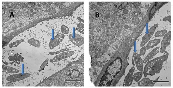
Figure 2.
Electron photomicrography of blood clots in mouse brains. A. Platelets (arrows) were mainly present in clots in small-diameter blood vessels (< 6 μm) after local clot formation. B. Platelets were also present in clots and accumulated near the endothelial wall in larger vessels (> 50 μm). Fibrin fibrils are visible in the clot. Scale bars, 2 μm.

