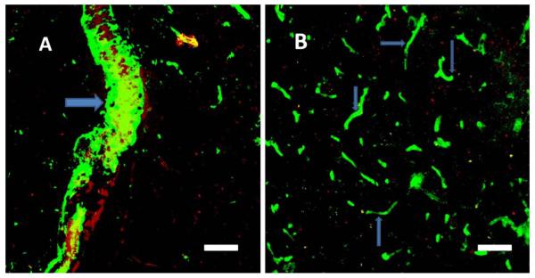
Figure 3.
Immunostaining of Aβ peptide after clot formation in blood vessels of the V2 cortex. In the visual cortex, (A) in the large vessel (> 50 μm, arrow) and (B) in small vessels (< 6 μm, arrows) Aβ peptide immunofluorescence was mainly concentrated in the vessel lumen. Erythrocyte-adsorbing red dye (Rose Bengal) was used for photothrombosis; thus, erythrocytes in the clots became visible in larger vessels. Scale bars, 40 μm.

