Figure 3.
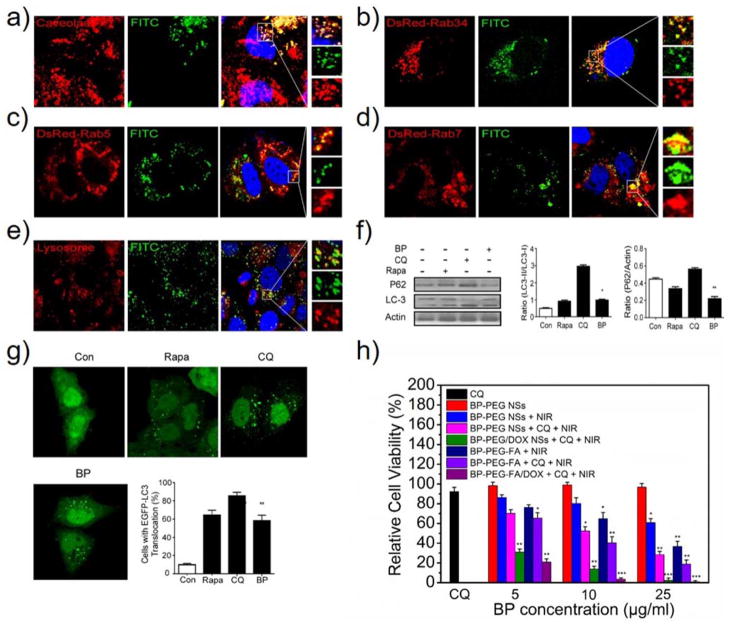
Endocytosis pathways and biological activities of PEGylated BP NSs. (a) CLSM images of HeLa cells incubated with BP-PEG-FITC NSs for 4 h, while caveolae were detected with primary antibodies against caveolae. CLSM images of HeLa cells transfected with (b) DsRed-Rab34, (c) DsRed-Rab5 and (d) DsRed-Rab7 after 4 h of incubation with BP-PEG-FITC NSs. (e) For lysosome detection, the HeLa cells were treated with BP-PEG-FITC NSs for 4 h and then were treated with Lyso-Tracker probes for 30 min. (f) HeLa cells were treated with BP-PEG NSs for 24 h, and then the LC3I/II and P62 protein levels were analyzed by western blotting. Histograms represent the quantitative analysis of LC3 and P62 protein expression performed by Image J, respectively (* P < 0.05, ** P < 0.01). (g) EGFP-LC3-transfected HeLa cells were treated with BP-PEG NSs for 24 h, and then quantification of cells with EGFP-LC3 vesicles was performed (** P < 0.01). (h) Relative viabilities of HeLa cells after different types of treatment at different BP concentrations (5, 10, 25 μg/ml). HeLa cells treated with only BP-PEG NSs or CQ (30 μM) was used as control (* P < 0.05, ** P < 0.01, *** P<0.001).

