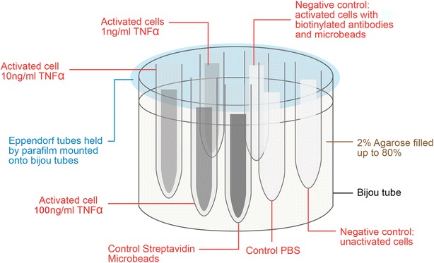
Fig. 2.

Cell phantom preparation. Six samples were prepared. They are the activated cells stimulated by TNF-α of different concentration (1 ng/mL, 10 and 100 ng/mL). The controls are activated cells with biotinylated IgG antibodies and microbeads as negative control, unactivated cells as negative control, and streptavidin microbeads
