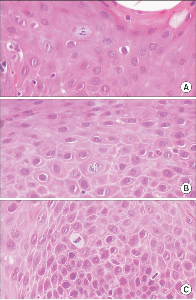
Fig. 3. A sample microscopic view of focal epithelial hyperplasia. There are a few mitosoid cells among the normal keratinocytes and chromatin peripheralization with inclusion bodies in the stratum granulosum (A), stratum spinosum (B), and stratum basale (C) (original magnification, ×400).