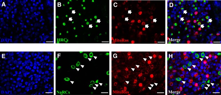
Fig. 10.
Differential staining of MitoTracker Red CMXRos in HRCs and NaRCs. A–D: fluorescent immunohistochemistry and confocal microscopy showing the staining of MitoTracker Red CMXRos (MitoRos) in H+-ATPase-rich cells (HRCs; depicted by the arrows). E–H: relatively weak staining of MitoRos in Na+-K+-ATPase-rich cells (NaRCs) was observed (depicted by the arrowheads). Nuclei were labeled by DAPI. HRCs and NaRCs were labeled by H+-ATPase and Na+-K+-ATPase antibody, respectively. Scale bar = 25 μm.

