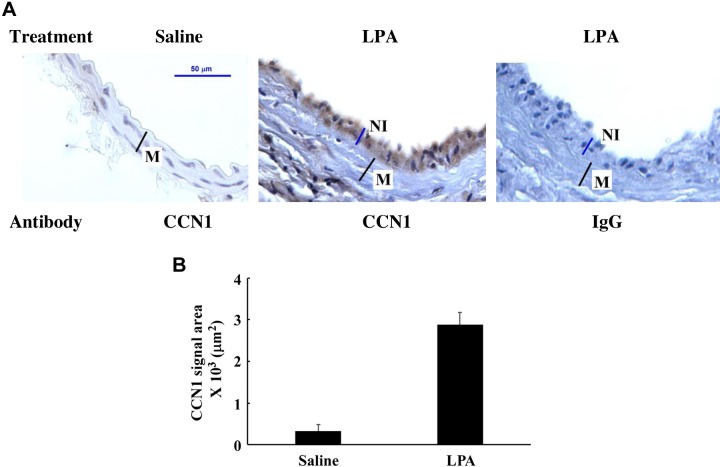
Fig. 4.
LPA induction of CCN1 expression in mouse CCA. CCA sections from LPA-infused (middle and right) or saline-infused (left) mouse carotid arteries were immunostained either with Cyr61 antibody (left and middle) or negative control, rabbit IgG (right). Nuclei were counterstained with hematoxylin. A: representative image of CCN1 expression detected by immunohistochemistry. B: graph illustration of detected CCN1 signal area. Values are means ± SD, n = 5 mice.

