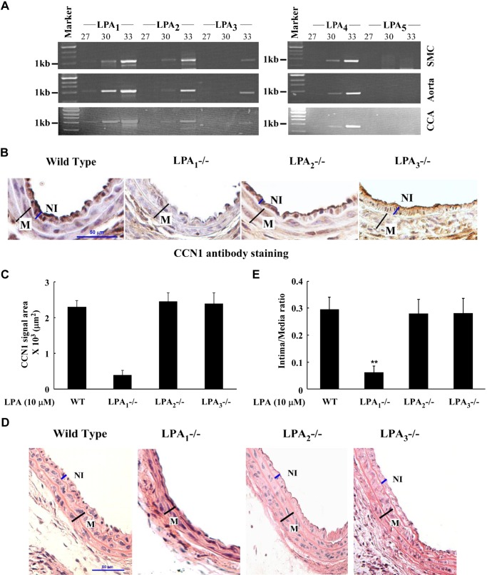
Fig. 6.
LPA receptor expression in mouse carotid arteries and effects of LPA receptor deficiency on CCN1 expression and neointimal formation. A: expression profiles of LPA receptors in mouse SMC, aorta, and CCA were detected by RT-PCR in various amplification cycles labeled at the top of each lane. B: expression of CCN1 in CCA of WT, LPA1−/−, LPA2−/−, and LPA3−/− mice infused with LPA. WT, LPA1−/−, LPA2−/−, and LPA3−/− mice were used for LPA infusion, and CCAs were harvested in 2 wk. CCN1 expression was examined with immunohistochemical staining. C: graph illustration of CCN1 in vivo expression. Immunohistochemical staining area was measured and compared between WT, LPA1−/−, LPA2−/−, and LPA3−/− groups. Values are means ± SD, n = 5 mice. D: neointimal formation in CCAs of WT, LPA1−/−, LPA2−/−, and LPA3−/− mice infused with LPA was detected by H and E staining. E: intima/media area was measured, and a graph illustration is shown. Data of all groups were analyzed using ANOVA, and comparisons between groups were performed using Dunnett's t-test. Values are means ± SD; five sections were analyzed in each mouse CCA; n = 7 mice. **P < 0.01 vs. WT group.

