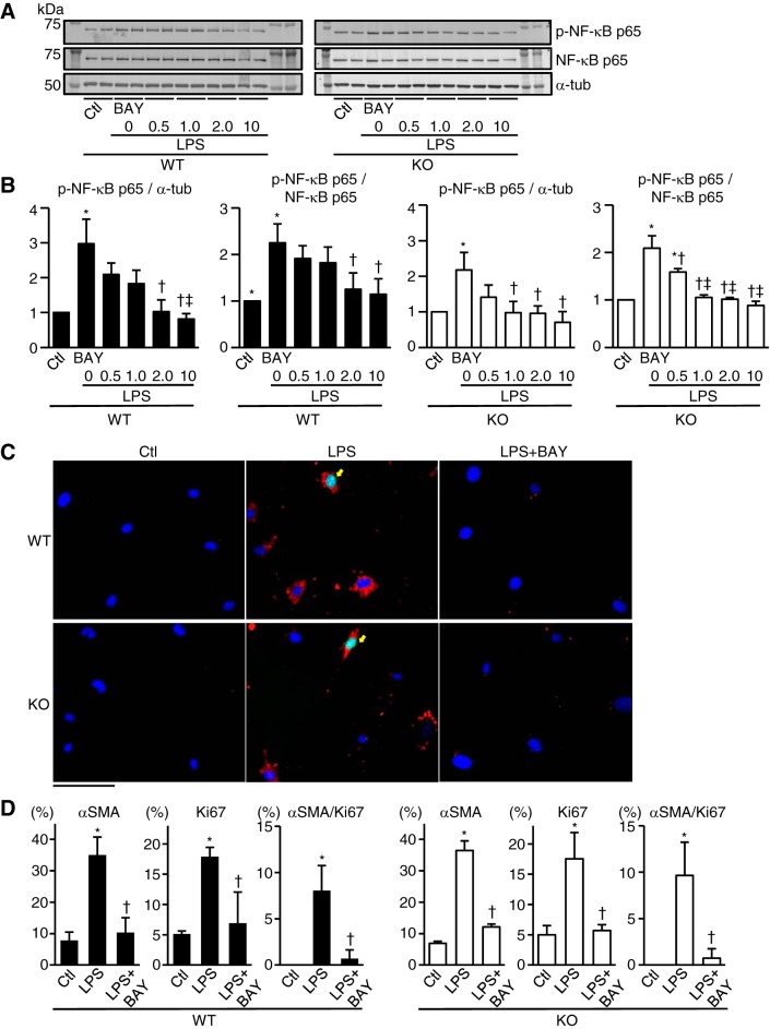
Fig. 8.
NF-κB activation and prevalence of α-smooth muscle actin (αSMA) and/or Ki67-positive cells in WT and TLR9-KO (KO) cardiac fibroblasts stimulated with lipopolysaccharides (LPS). A: representative Western blotting images. α-Tubulin (α-tub), NF-κB p65, and phospho-NF-κB p65 expressions were evaluated at 24 h after incubation with LPS (0.1 ng/ml) and BAY11-7082 (BAY0.5: 0.5 μM, BAY1.0: 1.0 μM, BAY2.0: 2.0 μM, BAY10: 10 μM). Control (Ctl) was incubated with medium without LPS or BAY11-7082. B: quantitative analyses of each protein expression level (n = 3 in each group). C: typical images of immunocytochemical stain. αSMA (red) and Ki67 (green) expressions were evaluated 48 h after incubation with LPS (0.1 ng/ml) and BAY11-7082 (2.0 μM). DAPI-positive nuclei are expressed in blue. Yellow arrows indicate αSMA and Ki67 double-positive cells. Scale bar = 100 μm. D: quantitative analyses of the percentage of αSMA-positive, Ki67-positive, and αSMA/Ki67-double-positive cells per DAPI-positive cells (n = 3 in each group). Data were analyzed by one-way ANOVA and are expressed as means ± SE. *P < 0.05 vs Ctl. †P < 0.05 vs LPS. ‡P < 0.05 vs LPS + BAY0.5.

