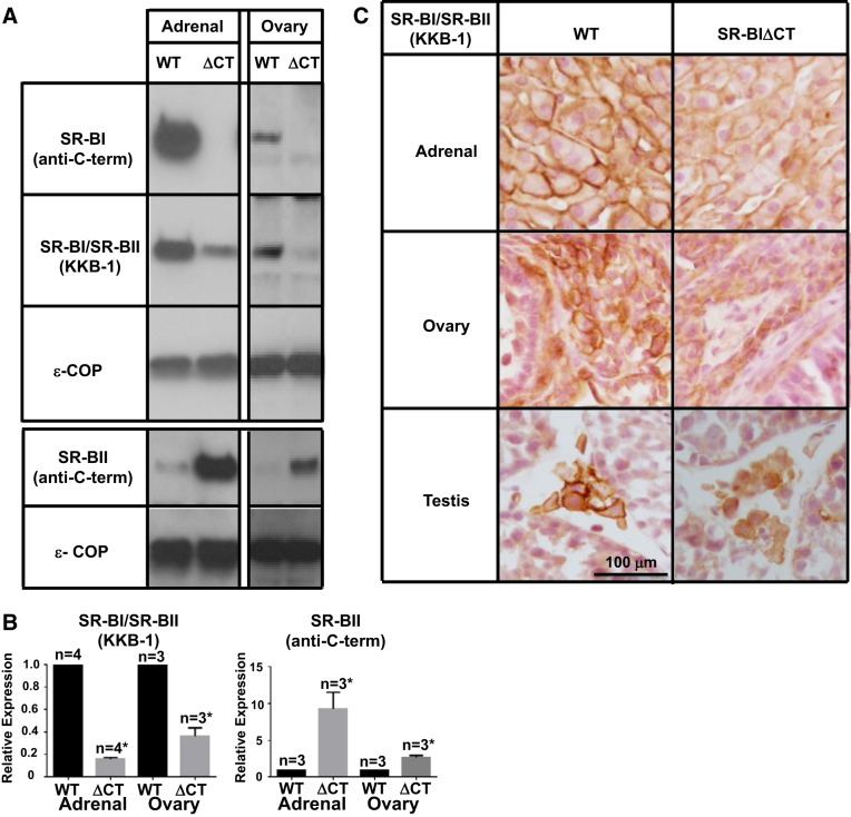
Fig. 3.
Effects of the SR-BIΔCT mutation on the expression of SR-BI and SR-BII proteins in steroidogenic tissues. A: immunoblotting analyses of adrenal gland and ovary lysates (∼30 μg protein) from WT and SR-BIΔCT 6- to 10-wk-old mice. Bands were visualized by chemiluminescence but with exposure times significantly shorter than those used in Fig 2A because of the higher protein expression in these steroidogenic tissues than in the liver. Proteins were detected with an SR-BI-specific [SR-BI (anti-C-term)] antibody, an anti-SR-BI/SR-BII (KKB-1) antibody, or an SR-BII-specific [SR-BII (anti-C-term)] antibody, as described in Fig 2. An anti-ε-COP antibody was used as a loading control. B: relative expression of SR-BI/SR-BII (KKB-1) and SR-BII in WT and SR-BIΔCT (ΔCT) mouse adrenal gland and ovary. *Significantly different [P < 0.0001 in adrenal glands and P = 0.0009 in ovaries for SR-BI/SR-BII (KKB-1) and P = 0.02 in adrenal glands and P = 0.003 in ovaries for SR-BII]. C: adrenal glands, ovaries, and testes from WT and SR-BIΔCT mice were fixed, frozen, and sectioned, and the sections were stained with the polyclonal anti-SR-BI/SR-BII KKB-1 antibody and a biotinylated anti-rabbit IgG secondary antibody and visualized by immunoperoxidase staining. Results for male and female mouse adrenal glands were identical. Magnification ×300.

