Fig. 5.
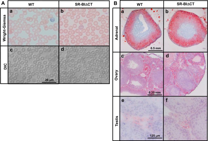
Red blood cell morphology (A) and Oil Red O (neutral lipid) staining of steroidogenic tissues (B) in WT and SR-BIΔCT mice. A: blood samples from 6- to 10-wk-old WT (a and c) and SR-BIΔCT (b and d) mice were stained with Wright-Giemsa and visualized by standard light microscopy (a and b) or differential interference contrast (DIC) optics (c and d). Magnification ×1,000. B: adrenal, ovarian, and testicular tissues from WT and SR-BIΔCT mice were frozen, and frozen sections (5 μm) were stained with Oil Red O-hematoxylin. Neutral lipids (e.g., cholesteryl esters) stain red. Representative sections are shown, and results for male and female mouse adrenal glands were essentially identical. Magnification ×25 adrenal gland, ×50 ovary, and ×250 testis.

