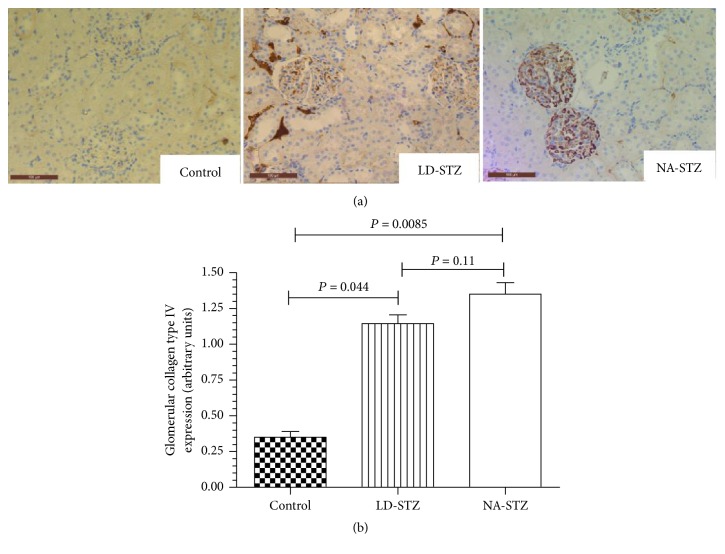
Figure 13.
Immunostaining of kidney cortex sections from control and diabetic rats with anti-type IV collagen antibodies. (a) Representative microphotographs from the experimental groups (magnification ×320). The markedly increased immunostaining is present in glomerular basement membranes and mesangial matrix of both diabetic groups as compared to control nondiabetic rats. (b) Comparison of the quantified staining intensity in glomeruli of the studied groups. Expression of type IV collagen was assessed semiquantitatively. Mean values and standard deviations are shown.

