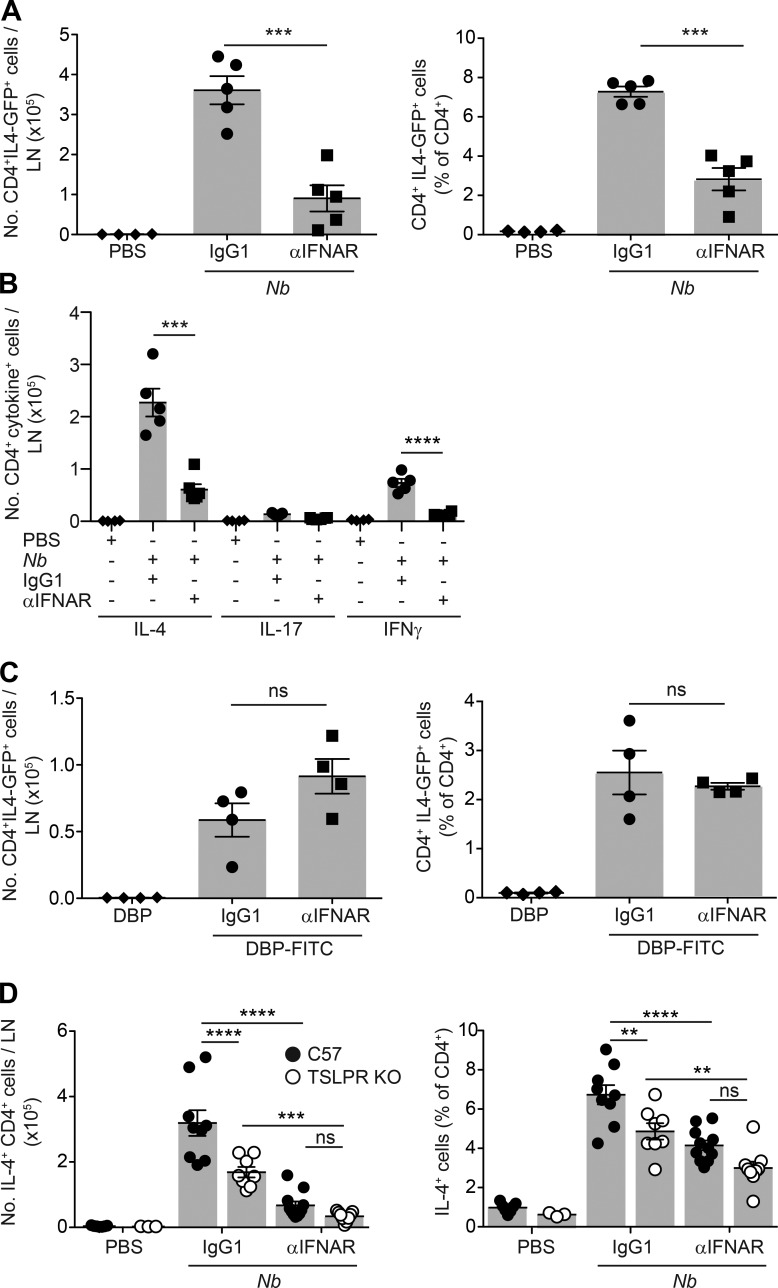
Figure 8.
IFN-I signaling is required for optimal priming of IL-4–producing T cells after Nb immunization and cooperates with TSLP in promoting Th2 responses. IL-4-GFP+/−, C57BL/6, or TSLPR KO mice were treated with 300 Nb, DBP-FITC, or the respective controls coadministered with αIFNAR or IgG1 isotype as indicated. 1 wk later, dLN were harvested for cytokine production assessment. (A) Numbers and percentages of IL-4-GFP+ CD4+ T cells in dLN as measured in IL-4-GFP+/− mice on day 7 after treatment with Nb and αIFNAR or IgG1 isotype control. (B) As in A, except that the number of cytokine+ T cells was measured in C57BL/6 mice by intracellular staining after in vitro restimulation with PMA and ionomycin. (C) As in A, except that IL-4-GFP mice were treated with DBP-FITC or DBP only. (D) As in A, except that the number of cytokine+ T cells was measured in C57BL/6 or TSLPR KO mice by intracellular staining after in vitro restimulation with PMA and ionomycin. Bar graphs show mean ± SEM for 3–11 mice/group; each symbol corresponds to one mouse. A–C show data from one of at least two repeat experiments that gave similar results. D shows combined data from two repeat experiments. P-values were determined using one-way (A–C) or two-way (D) ANOVA with the Bonferroni post-test; **, P < 0.01; ***, P < 0.001; ****, P < 0.0001; ns, not significant.

