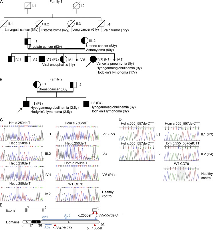
Figure 1.
Identification of CD70 mutations in two families with immunodeficiency and malignancy. (A) Family 1 pedigree. Cancer type, age of cancer diagnosis, and other key clinical manifestations in family members as well as the proband are shown. (B) Family 2 pedigree. Key clinical manifestations are shown. (A and B) /, deceased; double line, consanguinity; arrow, the proband (IV.6 in family 1 and II.1 in family 2); half-shaded, heterozygous; shaded, homozygous; unshaded, unknown; IV.5 and IV.7, miscarried fetus. (C) Sanger sequencing analysis of the CD70 gene in family 1. A homozygous (Hom) mutation was confirmed in the proband (P1) and the affected sibling (P2; IV.3). Heterozygous (Het) mutations were identified in the parents and three healthy siblings. (D) Sanger sequencing analysis of the CD70 gene in family 2. A homozygous mutation was confirmed in the proband (P3) and the affected sibling (P4). Heterozygous mutations were identified in the parents. Gray shading on the WT sequence indicates the bases that are deleted in the familial mutation. The sequences in this figure are in reverse direction. (C and D) The Sanger sequencing presented has been confirmed in at least two independent experiments. (E) The location of the mutation identified in the families in relation to the exon and protein domains (red triangles). The locations of antibodies (Ab1 recognizing amino acids 45–193, Ab2 recognizing amino acids 61–75, and Ab3 recognizing amino acids 129–193) used to detect CD70 protein expression are marked by blue arrows. C, cytoplasmic; E, extracellular; T, transmembrane.

