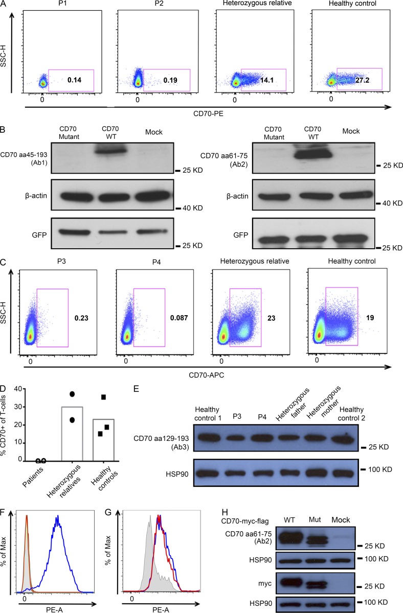
Figure 2.
Expression analysis of the CD70 mutants. (A) Expression of CD70 by peripheral blood B cells from the CD70-deficient patients (P1 and P2), compared with a heterozygous relative (father) and a normal control. CD70 expression was analyzed on CD19+-gated, live, single B cells. (B) Immunoblotting analysis of CD70 WT and mutant (identified in family 1) transfected HEK 293 cells. CD70 expression was probed with two different Abs specific for different epitopes (Ab1 and Ab2). GFP expression confirmed successful transfection, and β-actin served as a loading control. (C–E) Expression of CD70 by activated peripheral blood T cells from the two homozygous CD70-mutant patients (P3 and P4), compared with heterozygous parents (n = 2) and normal controls (n = 3). CD70 expression was analyzed by flow cytometry on CD3+ gated, live, single T cells using a mAb raised against CD70-transfected L cells (C and D) or by immunoblotting using polyclonal Abs (Ab3) raised against an extended C-terminal portion of CD70 (E). HSP90 was used to assess protein loading. (F) Binding of overexpressed WT or mutant CD70 to recombinant human CD27 was evaluated by flow cytometry. Shaded areas represent nontransfected 293T cells. Binding of anti-CD5 mAbs to cells transfected with WT (turquoise) or mutant (yellow) CD70 is shown. Binding of biotinylated CD27/streptavidin-PE in cells transfected with WT (blue) or mutant (red) CD70 is shown. (G) Flow cytometric detection of the flag epitope on cells transfected with WT (blue) or mutant (red) CD70 or on untransfected cells (shaded). (H) Immunoblotting of lysates from untransfected cells or cells transfected with WT or mutant CD70 plasmid, using Abs to CD70 (Ab2), myc tag, or HSP90. All data are representative of at least two independent experiments. Mut, mutant; SSC, side scatter.

