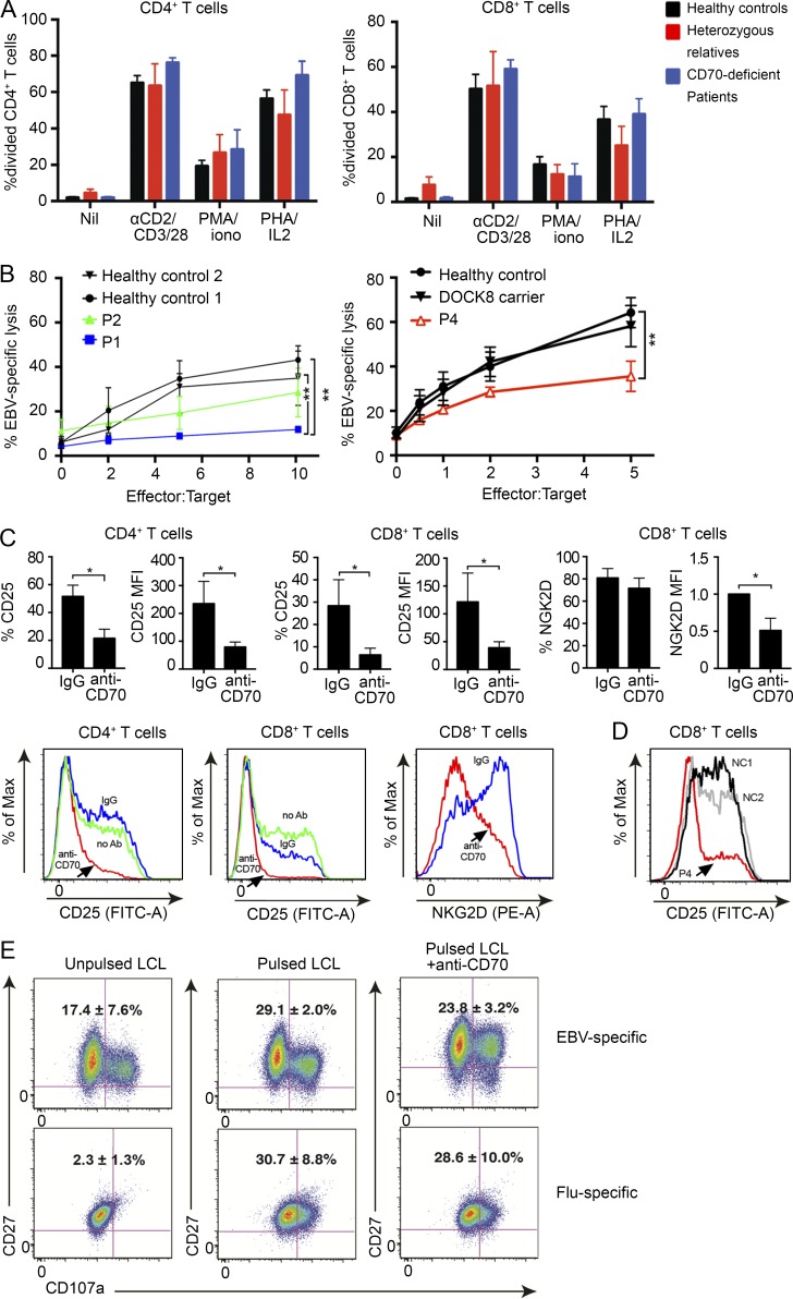
Figure 4.
Impaired cytotoxicity of CD70-deficient CD8+ T cells against EBV–B cell targets. (A) PBMCs from healthy donors (n = 11), heterozygous relatives (n = 3), and CD70-deficient patients (n = 4) were labeled with CFSE and then cultured in vitro in the absence (Nil) or presence of anti-CD2, CD3, CD28 beads, PMA/ionomycin (iono), or PHA/IL-2. Proliferation was determined after 4–5 d by determining the percentage of CD4+ or CD8+ T cells that had undergone one or more divisions. Values represent the mean ± SEM. (B) Percent lysis of autologous EBV-LCLs by EBV-specific CTL from P1 and P2 (compared with two healthy controls) and P4 (compared with a healthy control and a DOCK8+/− carrier). Shown are means ± SD from two (for P1 and P2) and four (for P4) experiments, respectively. **, P < 0.005 by one-way ANOVA. (C) Cell-surface activation marker expression on T cells, induced by EBV-LCLs in the presence of 10 µg/ml anti-CD70 or isotype control. Percent positive or geometric mean fluorescence intensity (MFI) of gated CD4+ T cells or CD8+ T cells, with CD25 measured at 4 d and NKG2D at 5 d of stimulation is shown. NKG2D mean fluorescence intensity was normalized to that of corresponding isotype control samples. Shown are representative histograms and means ± SD from treatments of PBMCs from four different healthy control donors without prior in vitro stimulation. *, P < 0.05 by a Mann-Whitney U test. (D) CD25 induction on T cells from two healthy controls or P4, after 4 d of stimulation by EBV-LCLs. Shown are representative histograms of gated CD8+ T cells during the fourth cycle of stimulation with irradiated autologous EBV-LCLs; similar results were obtained during the second or third cycles of stimulation. (E) EBV- or Flu-specific CD8+ T cell clones from healthy donors were cultured with autologous EBV-LCLs with or without specific peptides in the absence or presence of 10 µg/ml blocking anti-CD70 mAb. Expression of CD107a by the T cell clones was determined after 6 h. The values represent the mean ± SEM of two or three experiments using EBV-specific or Flu-specific clones, respectively. Note the increased level of activation of EBV-specific CD8+ T cell clones in the presence of unpulsed LCLs, over that observed for flu-specific clones, reflects the recognition of peptides by EBV-specific clones presented endogenously (i.e., without pulsing) by the autologous LCLs.

