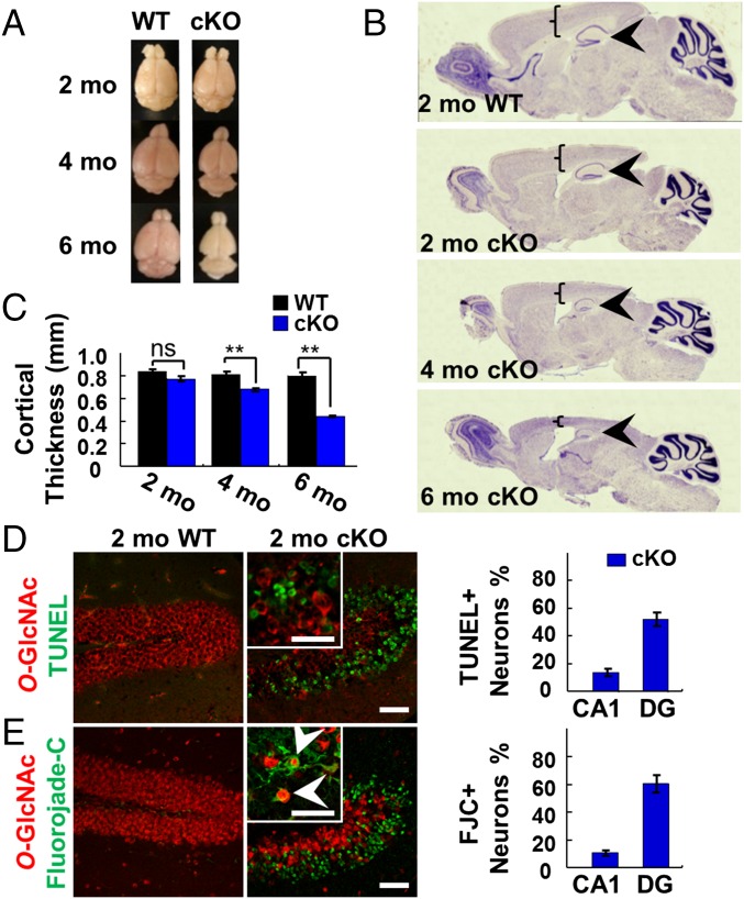
Fig. 2.
OGT cKO mice displayed reduced brain size and neurodegeneration. (A) Overall brain size was reduced at 4 and 6 mo in OGT cKO mice. (B and C) Nissl staining showed shrinking of cortical (brackets) and hippocampal (arrowheads) structures in OGT cKO mice (B) along with significant decreases in cortical thickness (C). (D) Costaining for O-GlcNAc (red) and TUNEL (green) identified apoptotic neurons in the hippocampus of OGT cKO mice. (E) Costaining for O-GlcNAc (red) and FJC (green) identified degenerating neurons in the hippocampus of OGT cKO mice, some of which were positive for O-GlcNAc (arrowheads). **P < 0.005. (Scale bars: 50 µm.)

