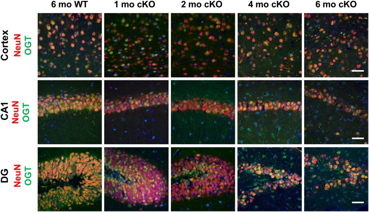
Fig. S1.
Time course for loss of OGT in the hippocampus and cortex of OGT cKO mice. An anti-OGT antibody was used to immunostain the cortex, CA1, and DG of OGT cKO and WT mice (green). NeuN was used to stain neuronal nuclei (red), and DAPI was used to stain DNA (blue). Loss of OGT expression began at 1 mo; by 4 mo most of the remaining neurons were OGT+ resulting from cell death of OGT-null neurons. Accordingly, progressive loss of neurons was also apparent at 4 and 6 mo. (Scale bars: 50 µm.)

