Fig. 4.
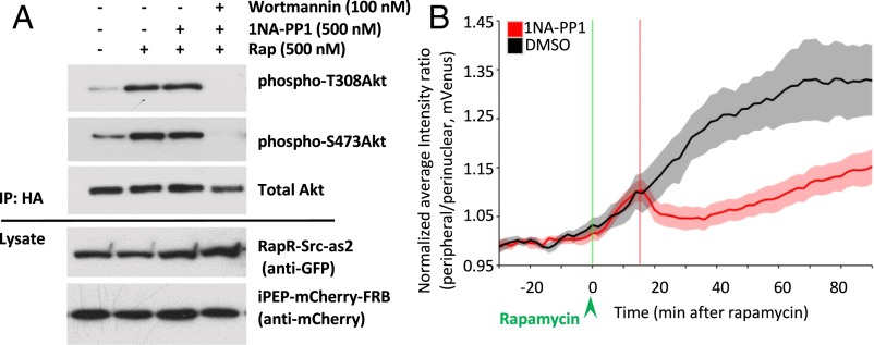
Regulation of PI3K signaling by RapR-Src-as2. (A) Akt phosphorylation. LinXE cells coexpressing cerulean-myc–tagged RapR-Src-as2, iPEP-mCherry-FRB, and HA-Akt were treated with rapamycin (500 nM) or ethanol (solvent) for 35 min and then with 1NA-PP1 (500 nM), 1NA-PP1 + Wortmannin (500 nM, 100 nM), or DMSO for 30 min. HA-Akt was immunoprecipitated and analyzed for phosphorylation at Thr308 and Ser473. Data are representative of three independent experiments. (B) Changes in peripheral/perinuclear mVenus-PH-AKT localization over time. HeLa cells, expressing RapR-Src-as2-cerulean-myc (adenovirus infection), mVenus-PH-AKT, and iPEP-mCherry-FRB (transfection) were imaged live. Cells were treated with rapamycin (500 nM) (green line), and 15 min later cells were treated with either DMSO (solvent) or 1NA-PP1 (250 nM) (red line). Shading represents 90% confidence intervals.

