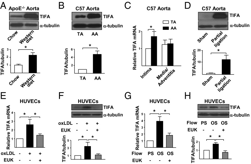
Fig. 1.
Oxidative stress activates TIFA in vivo and in vitro. (A) TAs were isolated from 8-wk-old ApoE−/− mice fed regular chow or Western diet for 8 wk. The TIFA expression in isolated tissue extracts was examined by Western blot analysis. (B and C) TA and AA were isolated from C57BL/6 mice. (B) Protein level of TIFA was examined by Western blot analysis. (C) Isolated vessels were further dissected to intima and media/adventitia portions, and the mRNA level of TIFA was quantified by RT-qPCR with primers listed in Table S1. (D) Partial ligated or sham control carotid arteries were dissected from C57BL/6 mice. Tissue extracts from three animals were pooled, and TIFA level was assessed by Western blot analysis. (E–H) HUVECs were pretreated with or without antioxidant EUK-134 (1 μM) for 2 h and then oxLDL (100 μg/mL), PS (12 ± 4 dyn/cm2), or OS (0.5 ± 4 dyn/cm2) for 16 h before cell lysate collection. TIFA mRNA level (E and G) and protein level (F and H) in cells were assessed accordingly. Data are mean ± SEM from three independent experiments (*P < 0.05).

