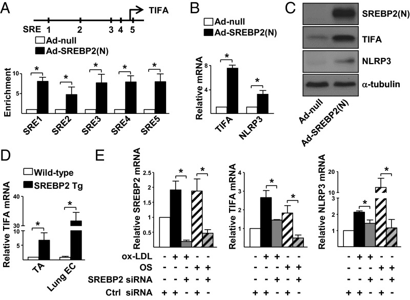
Fig. 2.
SREBP2 transcriptionally up-regulates TIFA in ECs. (A) HUVECs were infected with Ad-SREBP2(N) for 48 h. ChIP assays were performed by using anti-SREBP2 or rabbit IgG as an isotype control. The enrichment of SREBP2(N) binding to the putative SREs in the promoter region of the TIFA gene (Upper) was quantified by RT-qPCR with primers listed in Table S2. (B and C) HUVECs were infected with Ad-SREBP2(N) or Ad-null. The mRNA and protein levels of TIFA were examined by RT-qPCR and Western blot, respectively. (D) TA and lung ECs were isolated from EC-SREBP2(N)-Tg mice and WT littermates. The levels of TIFA mRNA in the tissues were quantified by RT-qPCR. (E) HUVECs were transfected with SREBP2 or control siRNA (20 nM). The transfected cells were treated with or without oxLDL or OS. The levels of SREBP2, TIFA, and NLRP3 mRNA were examined by RT-qPCR. Data are mean ± SEM from three independent experiments (*P < 0.05 between compared groups).

