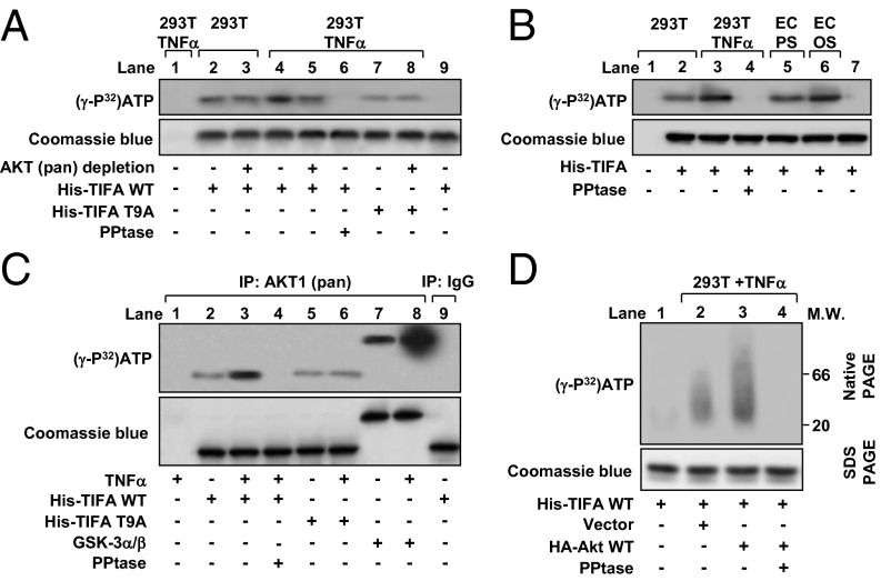
Fig. 4.
Akt is involved in the phosphorylation of TIFA Thr9. (A) HEK293T cells were treated with or without TNF-α for 30 min. Cell lysates were extracted, and Akt was depleted by immunoprecipitation (lanes 3, 5, and 8) before incubation with recombinant WT TIFA (His-TIFA-WT; lanes 2–6) or TIFA-T9A mutant (lanes 7 and 8) and [γ-32P]ATP. Alkaline phosphatase (PPtase) was added as indicated (lane 6). The reaction mixtures were separated by SDS-PAGE, and the bands were revealed by autoradiography. Lane 9 was a reaction mixture without cell lysates. (B) HUVECs were exposed to PS (lane 5) or OS (lane 6) for 30 min, and the collected cell lysates were incubated with recombinant WT TIFA and [γ-32P]ATP for in vitro kinase assay. Lysates from HEK293T cells treated with or without TNF-α (lanes 1–4) were used as positive controls, and lane 7 was reaction mixture without cell lysates. (C) Akt was immunoprecipitated from HEK293T cells treated with or without TNF-α for 30 min. The cell lysates were incubated with recombinant WT TIFA (lanes 2–4), TIFA-T9A mutant (lanes 5 and 6), or GSK-3α/β (lanes 7 and 8) together with [γ-32P]ATP for in vitro kinase assay. Rabbit IgG was an isotype control (lane 9). (D) HEK293T cells were transfected with or without the WT Akt and then treated with TNF-α for 30 min as indicated. His-TIFA-WT was incubated with cell lysates for in vitro kinase reaction. The polymerization of His-TIFA was analyzed by native gel electrophoresis. Coomassie blue staining was used for visualization and quantitation of TIFA protein in SDS-PAGE.

