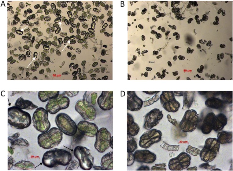
Fig. S8.
Microscopic evaluation of the enriched glandular secretory trichome extracts from A. annua WT leaves before and after boiling. Images were taken under bright field with 10× (A and B) or 40× (C and D) magnification for the intact trichome extracts (A and C) or 10-min boiled extracts (B and D). Intact subapical cavities are indicted by arrows (A and C). Scale bars are depicted in micrometers.

