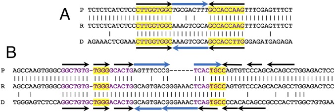
Fig. 3.
Examples of microinversions at predicted hairpins identified in cancer tissue from human individuals. Black arrows indicate the inverted repeats (IR), and blue arrows indicate the loop orientation. Other color codings are the same as in Fig. 1 B–E. All potential SPDIR events found in the human genomes are listed in Dataset S3. (A) The class 2 microinversion Z441, located in the protooncogene SASH1 of colon cancer tissue from individual 1. (B) Example of a microinversion that was fully annealed at the left IR but misannealed at the right IR (using an alternative microhomology for the right illegitimate joint), resulting in a net gain of six bp (Z2579; individual 3, colon cancer, intergenic region).

