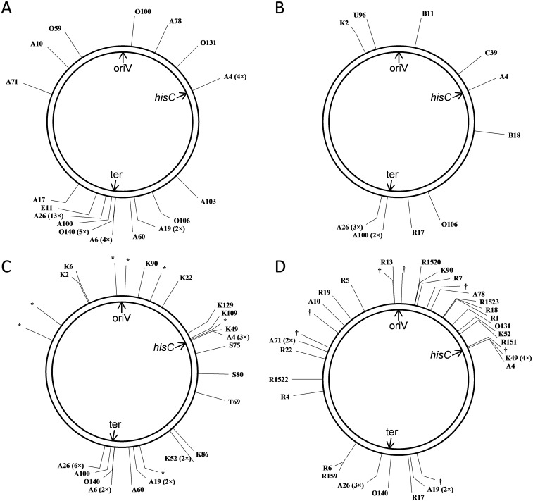
Fig. S1.
Schematic genomic positions of intragenomic donor DNA segments discovered in A. baylyi HisC+ SPDIR isolates. Numbers in brackets indicate the number of findings in independent experiments. The circles represent the A. baylyi chromosome (3.6 Mbp). oriV, origin of replication; ter, terminus of replication (approximate position); hisC, location of the hisC::′ND5i′ detection construct. All recombinant sequences are shown in Dataset S1. (A) Positions in recA+ strains (WT, ΔrecJ, ΔexoX, ΔrecJ ΔexoX, and ΔcomA ΔrecJ ΔexoX) without genotoxic stress or added DNA. (B) Positions in recA+ (WT and ΔrecJ ΔexoX) strains after exposure to ciprofloxacin or UV light. (C) Positions in recA+ strains without stress after addition of DNA (isolated from B. subtilis or salmon sperm or from isogenic A. baylyi hisC::′ND5i′ DNA). The donor DNA loci for K143 (Dataset S1) are identical with sequences from 23S rRNA genes of both B. subtilis (10 copies per genome) and A. baylyi (7 copies), and the A. baylyi positions are indicated with an asterisk. (D) Positions in ΔrecA strains (recJ+ exoX+ and ΔrecJ ΔexoX). The donor DNA for K1518 (Dataset S1) originates from one of the seven 16S rRNA genes (positions indicated by a dagger). Designed with pDRAW32 (www.acaclone.com).

