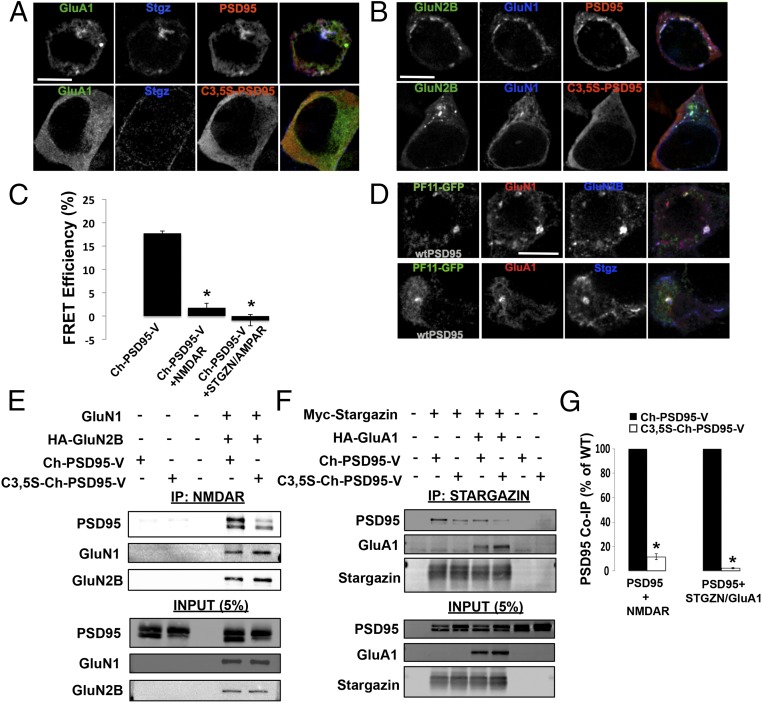
Fig. 2.
Palmitoylation of PSD95 regulates interactions with AMPARs and NMDARs. (A) Immunolocalization of HA-Stargazin, GFP-GluA1, and untagged wild-type (wt) PSD95 or C3,5S-PSD95 in HEK293 cells. (Top) PSD95, HA-Stargazin, and GFP-GluA1 AMPAR subunits transfected in HEK293 cells. (Bottom) C3,5S-PSD95 replaced PSD95. In both sets of images, PSD95 or C3,5S-PSD95 was detected with anti-PSD95 antibodies, and with anti-HA antibodies for Stargazin. (Scale bar: 10 μm.) (B) Immunolocalization of NMDARs (GFP-GluN2B and Flag-GluN1) and untagged PSD95 or C3,5S-PSD95 in HEK293 cells. (Top) PSD95, GFP-GluN2B, and Flag-GluN1 NMDAR subunits transfected in HEK293 cells. (Bottom) C3,5S-PSD95 replaced PSD95. In both sets of images, PSD95 or C3,5S-PSD95 was detected with anti-PSD95 antibodies, and with anti-Flag antibodies for GluN1. (Scale bar: 10 μm.) (C) Ch-PSD95-V FRET analysis to test for conformational changes that occur with interactions with NMDARs and AMPARs in HEK293 cells. Cells were transfected with Ch-PSD95-V and NMDARs, consisting of HA-GluN2B/Flag-GluN1, or with AMPARs, consisting of Myc-Stargazin/HA-GluA1. (D) Palmitoylated PSD95 specifically colocalized in puncta with NMDARs and AMPARs in HEK293 cells. Cells were transfected with PF11-GFP, the conformation-specific intrabody for palmitoylated PSD95, untagged wtPSD95, and either Flag-GluN1 plus HA-GluN2B (NMDARs, Top) or mCherry-GluA1 plus HA-Stargazin (AMPARs, Bottom). Flag-GluN1, HA-GluN2B, and HA-Stargazin were detected with anti-Flag and anti-HA antibodies. (Scale bar: 10 μm.) (E) Loss of PSD95 palmitoylation sites disrupts interactions between PSD95 and NMDARs. PSD95–NMDAR interactions were assayed by co-IP and Western blots. A representative Western blot shows the levels of Ch-PSD95-V that coimmunoprecipitated with GluN2B subunits. Cell lysates prepared from HEK293 cells expressing NMDARs (Flag-GluN1 and HA-GluN2B) were mixed with separate lysates from cells expressing Ch-PSD95-V or C3,5S-Ch-PSD95-V. IPs were performed with anti-HA antibodies specific for GluN2B, and anti-PSD95 and anti-HA antibodies were used to blot. (F) Loss of PSD95 palmitoylation sites disrupts interactions between PSD95 and AMPARs containing Stargazin. A representative Western blot displays the levels of Ch-PSD95-V that coimmunoprecipitated with Stargazin subunits alone, or with Stargazin and GluA1 subunits. Cell lysates prepared from HEK293 cells expressing Stargazin-containing AMPARs (Myc-Stargazin and HA-GluA1) or Stargazin alone (Myc-Stargazin) were mixed with separate lysates from cells expressing Ch-PSD95-V or C3,5S-Ch-PSD95-V. IPs were performed with anti-Myc antibodies specific for Stargazin, and anti-PSD95 and anti-HA antibodies were used to blot. (G, Left) Quantification of band intensities for Ch-PSD95-V and C3,5S-Ch-PSD95-V that coprecipitated with NMDAR subunits from four separate Western blots. Band intensities are plotted as the percentage of wild-type (WT) PSD95, with Ch-PSD95-V set at 100%. The mean value for C3,5S-Ch-PSD95-V band intensities was 11.6 ± 2.3% (SEM), with *P < 0.001. (G, Right) Quantification of band intensities for Ch-PSD95-V and C3,5S-Ch-PSD95-V that coprecipitated with AMPAR subunits from four separate Western blots. Band intensities are plotted as the percent of WT PSD95, with Ch-PSD95-V set at 100%. The mean value for C3,5S-Ch-PSD95-V band intensities was 2.23 ± 0.5% (SEM), with *P < 0.001.

