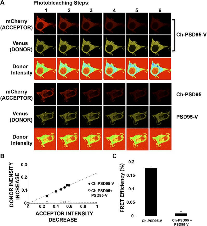
Fig. S2.
Ch-PSD95-V FRET in HEK293 cells. (A) Representative images of the photobleaching method performed on Ch-PSD95-V in HEK293 cells to obtain Ch-PSD95-V intramolecular FRET efficiency (Top). Venus and mCherry fluorescence images were acquired before and after six acceptor photobleaching steps. Venus (donor) pseudocolor intensity is displayed in the third row. (Bottom) No rise in donor intensity is observed when Ch-PSD95 is coexpressed with PSD95-V. (B) Ch-PSD95-V FRET efficiency determined from the linear relationship between increases in donor (mCherry) fluorescence with decreases in acceptor (Venus) with photobleaching. The photobleaching steps for ChPSD95-V in A are plotted as the percent decrease in acceptor intensity versus the percent increase in donor intensity (●). A corresponding rise in donor intensity is not observed when Ch-PSD95 is coexpressed with PSD95-V (○). (C) FRET efficiency value, ET, was calculated from the data above using the equation: ET = (1 − Ida/Id), where Ida and Id are the fluorescence intensities of the donor in the presence and absence of acceptor, respectively. FRET efficiency for Ch-PSD95-V is 17.7 ± 0.4% (mean ± SEM; n = 27 cells, *P < 0.00001) compared with 0.9 ± 0.7% (mean ± SEM; n = 19 cells) in cells coexpressing Ch-PSD95 and PSD95-V.

