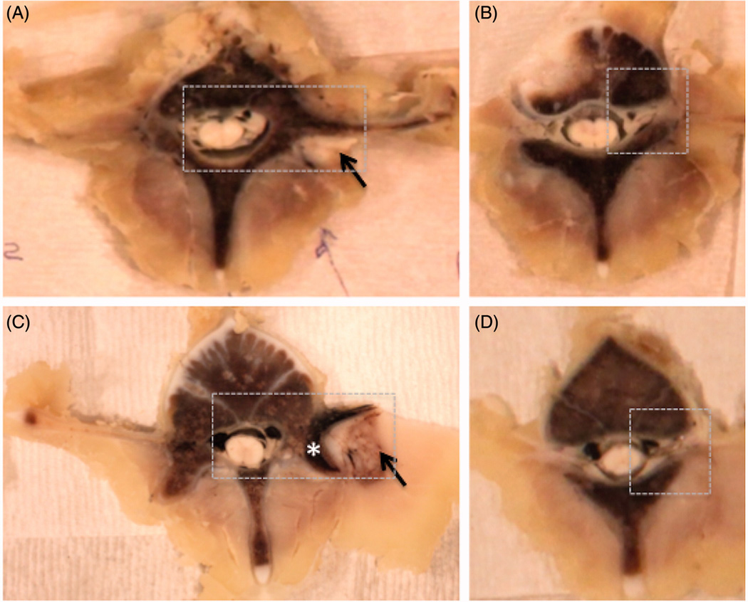
Figure 6.

Examples of gross pathology from the acute experiment 2 and subacute experiment 1. Ablation in muscle tissue (black arrow) appears as a paler area in the acute case (A-B) and the area surrounded by the brown rim in the subacute case (C–D). The brown rim of hemorrhage is apparent in the superficial region of the bone (asterisk) in the subacute case (C). The nerve roots, superior to the ablations, appear normal on gross pathology (B, D). The dashed box shows the region selected for histological analysis. Subacute gross pathology image corresponds to the follow-up images in Fig 4.
