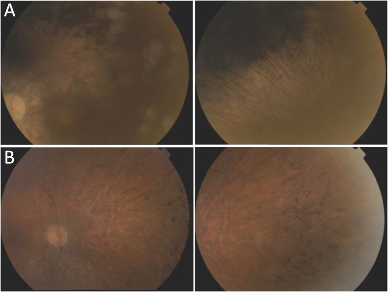
Figure 1. Fundus photographs of patients with novel mutations in PRPF8.
(A) Patient RP90 (p.Val2325_Glu2330del) shows optical disc pallor, arteriolar attenuation and macular atrophy (right), with dense pigment in the mid-periphery (left). (B) Patient RP148 (p.Leu2315Leufs*2336Aspext*21) shows optical disc pallor, arteriolar attenuation and bone spicule-shaped pigment deposits in the mid-periphery. The left and right pictures correspond to the left and right eyes, respectively.

