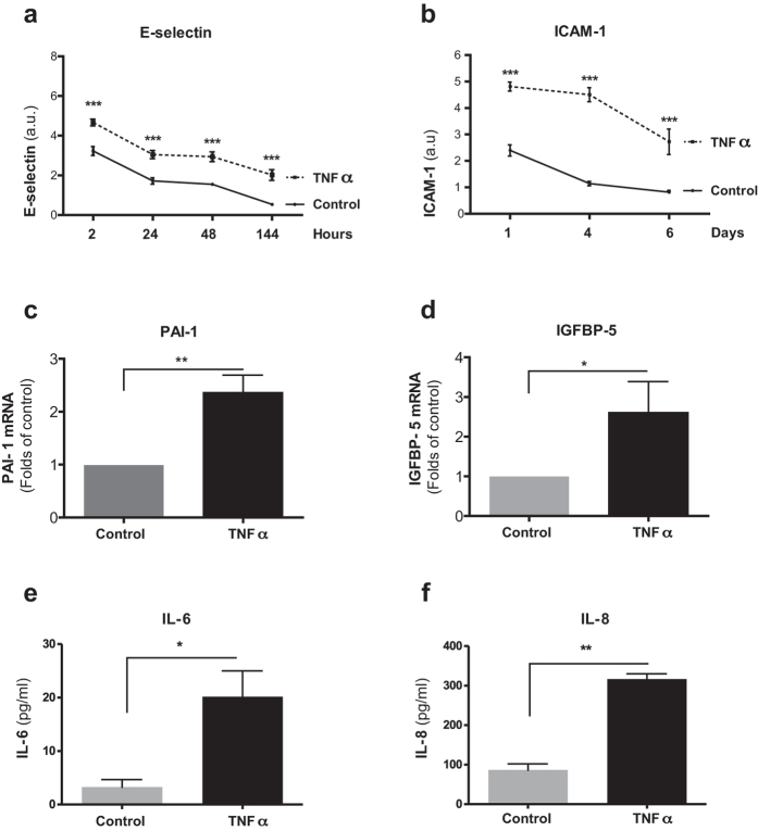
Figure 2. Prolonged TNFα-exposure promotes SASP in HUVECs.
(a,b) Surface expression of E-selectin (a) and ICAM-1 (b) of HUVECs grown in the presence of TNFα (10 ng/ml) in comparison to control HUVECs for indicated time points, quantified by cell ELISA. (c,d) mRNA levels of PAI-1 (c) and IGFBP-5 (d) of HUVECs grown in the presence of TNFα (10 ng/ml) in comparison to control HUVECs at day six, quantified by qPCR. Levels of (e) IL-6 and (f) IL-8 in supernatants of HUVECs grown in the presence of TNFα (10 ng/ml) in comparison to control HUVECs at day six, quantified by ELISA. Values are presented as mean ± SD of technical triplicates (a–d) or technical duplicates (e,f). (*p < 0.05, **p < 0.01, ***p < 0.001). a.u.: arbitrary units. The results shown are representative of three independent experiments.

