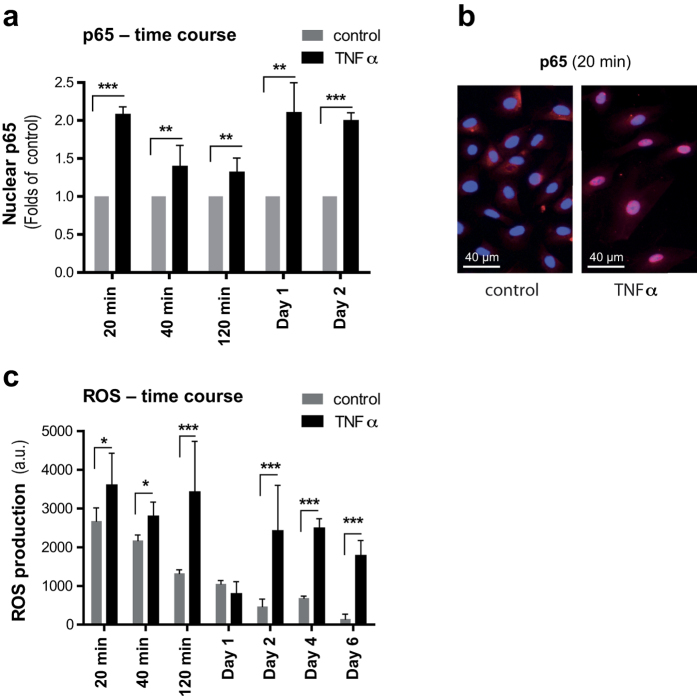
Figure 3. TNFα increases the nuclear translocation of NF-κB and induces ROS generation in HUVECs.
(a) Quantification of nuclear translocation of NF-κB in HUVECs grown in the presence of TNFα (10 ng/ml) for indicated time points, in comparison to control HUVECs, by integrated mean intensity of p65 in nucleus using CellProfiler software. (b) Representative fluorescent images of nuclear translocation of NF-κB in HUVECs (20 min) grown in presence or absence of TNFα. (c) Determination of ROS production in HUVECs grown in the presence of TNFα (10 ng/ml) for indicated time points, in comparison to control HUVECs, by using fluorophore H2-DCF (10 μM). Values are presented as mean ± SD of technical triplicates (*p < 0.05, **p < 0.01, ***p < 0.001). a.u.: arbitrary units. The results shown are representative of three independent experiments.

