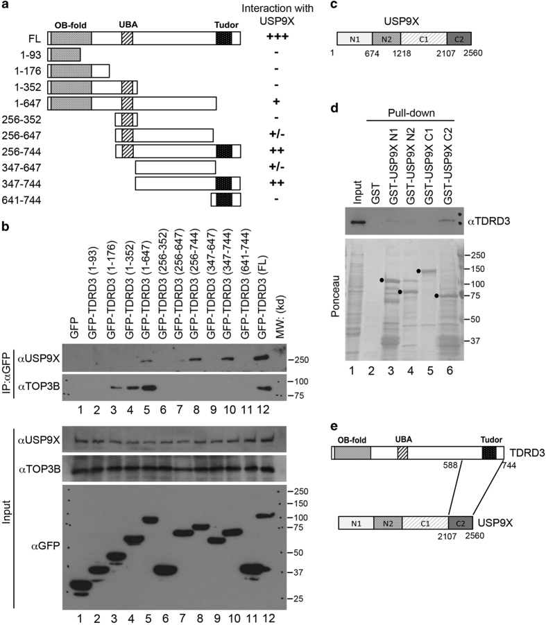
Figure 2.
Mapping the interaction regions of TDRD3 with USP9X. (a) A series of GFP-fusion deletions of TDRD3 were generated. The locations of the OB fold (OB-fold), the ubiquitin-binding domain (UBA) and the Tudor domain (Tudor) are indicated. A summary of the interactions observed in b is shown. (b) A co-IP assay was performed in HeLa cells transfected with the different TDRD3 GFP-fusion vectors. The cell lysates were IPed with anti-GFP, and the eluted samples were blotted with anti-USP9X and anti-TOP3B. The input samples were blotted with anti-USP9X, anti-TOP3B and anti-GFP. (c) A diagram indicates the GST-fusion deletions of USP9X generated for the pull-down assays described in d. (d) GST pull-down assays were performed using recombinant GST, GST-USP9X (N1), GST-USP9X (N2), GST-USP9X (C1) and GST-USP9X (C2) with the HeLa cell total cell lysates. Both the input samples and pull-down samples were detected with anti-TDRD3 (upper panel). The GST-tagged recombinant proteins in the pull-down samples were visualized by Ponceau S staining (bottom panel). (e) A graphical summary of the protein regions that mediate the TDRD3–USP9X interaction.

