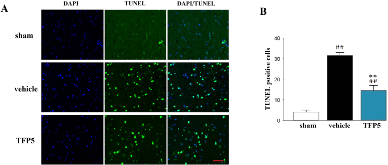
Figure 4. A dose of 30 mg/kg of TFP5 significantly reduced apoptosis in the ischemic regions of animals that underwent 2 h of MCAO and 48 h of blood reperfusion.
(A), Representative photomicrographs of immunofluorescence labeled with DAPI (blue) and apoptosis of brain cells (green) in the ischemic regions of animals at 48 h after 2 h-MCAO. Bar, 20 μm. (B), Semiquantitative results of apoptosis of brain cells. The bars represent means ± SEs. ##p < 0.01 versus sham group; **p < 0.01 versus vehicle group. MCAO, middle cerebral artery occlusion; DAPI, 4′,6-diamidino-2-phenylindole.

