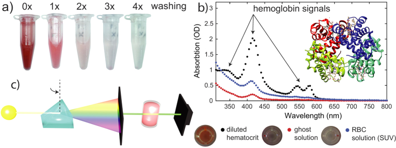
Figure 6. Removal of hemoglobin from the erythrocyte blood fraction after induced lysis in hypotonic buffer.
(a) Ghost samples lose their characteristic red color through sequential centrifugation and washes. (b) Comparison of UV-vis absorbance curves at different stages within ghost preparation. Characteristic hemoglobin absorbance signatures are significantly reduced in the final solution after the procedure. (c) Schematic of the UV-vis setup.

