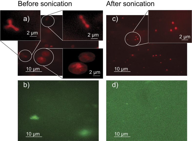
Figure 7. Fluorescence microscopy images of the ghost solution before and after sonication.
The membrane was labelled using DiI in parts (a) and (c), while Alexa Fluor 488 labelled phalloidin was used to label the F-actin network in (b) and (d). Before sonication, ghosts of highly irregular shape and a large size distribution are observed including ‘ghosts inside of ghosts’. The solution also contains large clusters of actin. Small vesicles with a uniform size distribution are observed after sonication and no actin particles (within the resolution of the microscope used).

