Fig. 3.
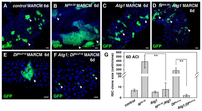
Tumor formation in the absence of Notch signaling can be effectively inhibited by induction of autophagy in the adult Drosophila midgut. (A) ISC MARCM clones (green) in FRT control 6 days after clone induction (6D ACI) (white arrowheads). (B) Progenitor tumors (green) are formed in N264-39 ISC clones (6D ACI) (white arrowheads). (C). ISC MARCM clones (green) expressing Atg1 (6D ACI) (white arrowheads). (D) Progenitor tumors observed in N264-39 ISC clones are significantly inhibited by co-expression of Atg1 (white arrowheads). (E) Progenitor tumors (green) are formed in DlRevF10 ISC clones (6D ACI) (white arrowhead). (F) Progenitor tumors observed in DlRevF10 ISC clones are almost completely inhibited by co-expression of Atg1 (white arrowheads). (G) Quantification of relative size of ISC clones (cell/clone) in the intestines of different genotypes. Note that both N264-39 and DlRevF10 ISC clones are highly deformed, making it difficult to accurately count the number of mutant cells. n=5–10 intestines. Mean ± SD is shown. **p<0.001. Blue indicates DAPI staining for DNA. Scale bars: 20 μm.

