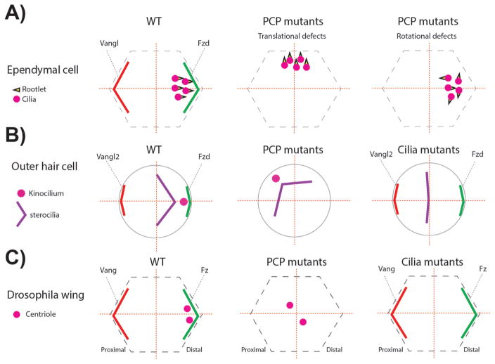
Figure 5.
PCP and cilia mutants associated phenotypes related to centriole/basal body (BB) A: In ependymal cells BBs are localized near the Fzd expression domain. In addition, the basal foot of the cilia, called rootlet and critical for ciliary beating, is aligned in the same direction (left panel). In one set of specific PCP mutants (check text for specific mutants) the localization of the BBs leading to so-called “translational defects” (middle panel). In other PCP mutants, although the translational polarity is correct, the alignment of the ciliary rootlets is uncoordinated (right panel). B: In the inner ear, outer hair cells (OHCs) position the BBs of the kinocilium (a specialized primary cilium) near the Fzd-Dvl localization domain, similar to ependymal cells (left panel). In PCP mutants the off-centered positioning of the BB is affected, but the V-shaped stereocilia bundle is not affected, following the position of the BB/kinocilium (middle panel). In cilia mutants, the V-shape of the stereocilia is affected and the structure remains central. The stereocilia bundle can appear as a line or circular shape depending on the severity of the ciliary mutant, in which the kinoclium is missing (right panel). C: Once fully polarized in Drosophila pupal wing cells, centrioles are localized near the Fz-Dsh domain, similar to mouse ependymal cells and OHCs (left panel). In PCP core and effector mutants, centrioles remain centered (middle panel). In Sas4 mutants where centrioles are absent, PCP remains mostly normal and actin polymerization in trichomes is largely unaffected (right panel).

