Figure 1.
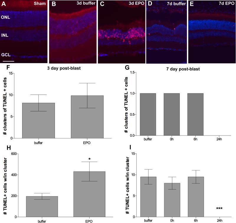
Treatment with EPO beginning at 24h post-blast decreases cell death at 7, but not 3-days post-blast. (A-E) Representative fluorescence micrographs of clusters of TUNEL-positive cells in retinas from: (A) sham blast, (B) buffer-injected 3-day post-blast, (C) EPO-injected 3-day post-blast, (D) buffer-injected 7-day post-blast, and (E) EPO-injected 7-day post-blast DBA/2J mice. TUNEL (red), DAPI (blue). Scale bar represents 50μm. (F, G) Bar graphs of clusters of TUNEL-positive cells at 3-days post-blast (F) and 7-days post-blast (G). (H, I) Bar graphs of the density of TUNEL-positive cells within each cluster at 3-days (H) and 7 days post-blast (I). Only mice injected with EPO at 24h post-blast and assessed at 7 days post-blast lacked TUNEL positive cells, ***p<0.001. Note that (G) only shows results from retinas that contained clusters of TUNEL cells, with the exception of the 24h group which contains data from all retinas since all were completely TUNEL negative. Only 18% of the buffer, 0h, and 6h 7-day post-blast retinas contained TUNEL-positive cells.

