Figure 2.

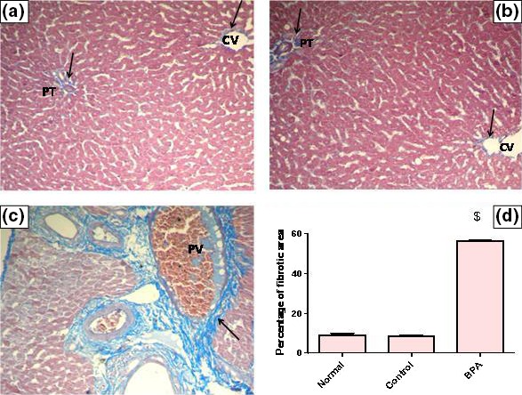
Representative photomicrographs of liver sections stained by Mallory trichrome (×200): (A) A liver section of an adult male albino rat of normal group, showing the normal distribution of collagen fibers around central vein (CV) and portal tract (PT) (arrows). N.B. The collagen fibers are stained blue. (B) A liver section obtained from an adult male albino rat of control group receiving vehicle (corn oil), showing normal distribution of collagen fibers around central vein (CV) and portal tract (PT) (arrows). (C) A liver section taken from an adult male albino rat receiving BPA, showing a marked increase in collagen fibers deposition around portal tract (arrow) and congested portal vein (PV) [Mallory trichrome × 200]. (D) The mean fibrosis (collagen) area percentage/μm2 surface area of liver tissue in the studied groups of adult male albino rats. Values were expressed as mean ± SD (n = 6/group). $ Significantly different from control group at P < 0.001 using anova followed by Tukey–Kramer as a post hoc test. BPA, bisphenol A.
