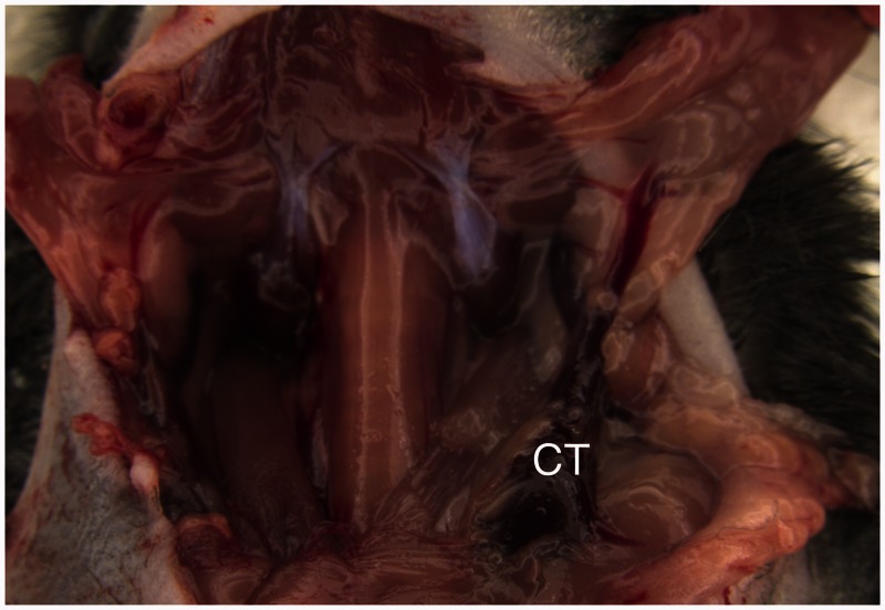
Figure 1.
Surgical exposure of the left external jugular vein (EJV) after dorsolateral reflection of the salivary gland. Note the large branch running from the salivary gland into the vein. CT: the common trunk of the EJV, which was selected for the blood flow interruption and subsequent division of the vein

