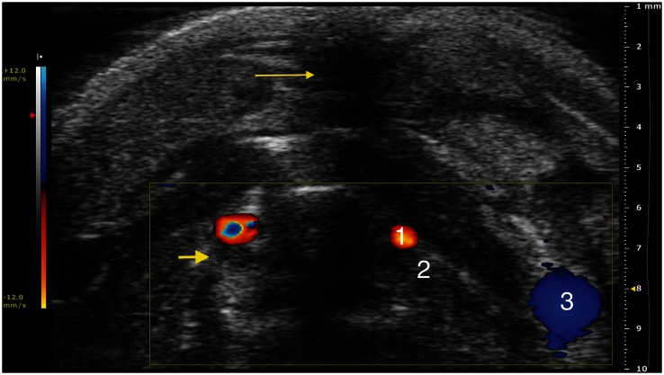
Figure 4.
Trans-axial color-Doppler HFUS image at a PRF of 1 kHz (12.0 mm/s blood velocity). (1) Carotid artery, (2) internal jugular vein, (3) external jugular vein. Note: Acoustic shadowing due to surgical access (thin arrow); moderately hyperechoic material is visible within the internal jugular vein, in addition to the absence of flow, which is compatible with thrombotic phenomena following the interruption of the blood flow (large arrow)

