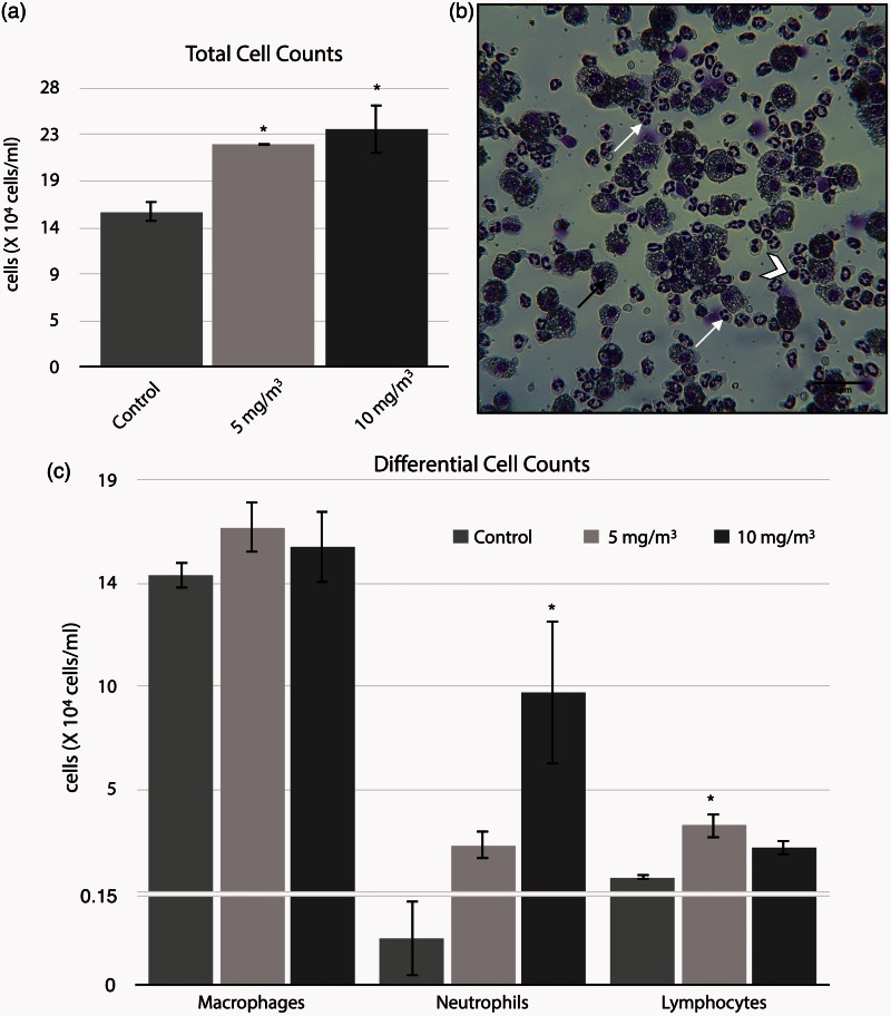
Figure 2.
Figure representing the concentration-response effect to WO3 NPs of markers for inflammation and cytotoxicity in the bronchoalveolar lavage collected from hamster lungs, 24 h post-inhalation. BAL fluids and cell were separated by centrifugation. The cells were re-suspended in 1 mL of RPMI 1640 media, followed by determination of total cell numbers (a) by direct counting with the use of a hemocytometer. The total cell numbers were significantly (P < 0.05) increased ∼1.5 folds in the treatment groups. (b) Photomicrograph of BALF cells from lung exposed to 10 mg/m3 of WO3 NPs for 4 h/day for four days. Note the presence of PMNs (arrow), lymphocyte (chevron), and macrophages (dark arrow). Differential counts (c), slide smears were prepared by using a cytocentrifuge showed neutrophilia by a statistically significant (P < 0.05), 27% increase in neutrophils (10 mg/m3 treated group) as compared to control. (A color version of this figure is available in the online journal.)

