
Figure 1.
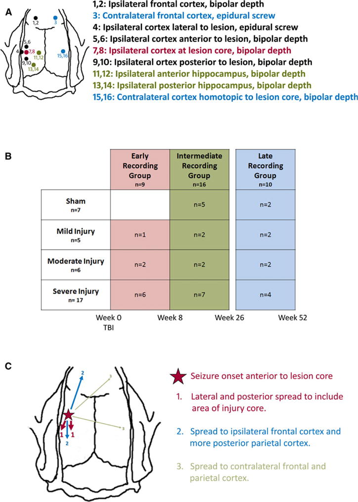
(A) Schematic representation of depth micro-electrode and epidural screw electrode placement for continuous EEG recordings after lateral fluid percussion injury. Electrodes denoted in red are within the lesion core, electrodes denoted in black are within the neocortex ipsilateral to the injury, electrodes denoted in green are within the ipsilateral hippocampus, and electrodes denoted in blue are in the hemisphere contralateral to the injury. (B) Schematic of the experimental design showing number of animals within each injury severity and each recording time. (C) Schematic showing the average spread pattern of a focal seizure. The seizure was first recorded by electrodes in the neocortex just anterior to the lesion site (red star), and then would usually spread first to neocortical electrodes in the lesion core and the area just lateral to it (areas marked 1). Next the seizure would usually spread to electrodes in the ipsilateral frontal neocortex and more posterior parietal neocortex (areas marked 2), and finally in some cases it would also spread to the contralateral frontal neocortex and area homotopic to the lesion (areas marked 3).
Epilepsia © ILAE