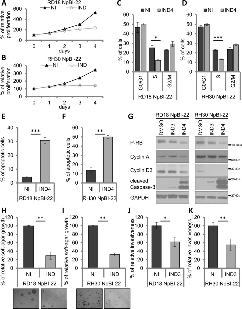
Figure 2.
miR-22 interferes with the transforming abilities of RMS cells. A and B, proliferation analysis of inducible miR-22-expressing RD18 (A) and RH30 (B) NpBI-22 cells (induced, IND; non-induced, NI). C and D, cell cycle distribution of RD18 (C) and RH30 (D) NpBI-22 cells (induced, IND; non-induced, NI). E and F, apoptosis assessment of RD18 (E) and RH30 (F) NpBI-22 cells (induced, IND; non-induced, NI). G, Western blot analysis in RD18 and RH30 NpBI-22 cells (induced, IND; non-induced, NI). H and I, quantification and representative images of soft-agar growth of RD18 (H) and RH30 (I) NpBI-22 cells (induced, IND; non-induced, NI). J and K, invasiveness assessment of RD18 (J) and RH30 (K) NpBI-22 cells (induced, IND; non-induced, NI). Error bars, SEM. *P<0.05; **P<0.01; ***P<0.001 (t test).

