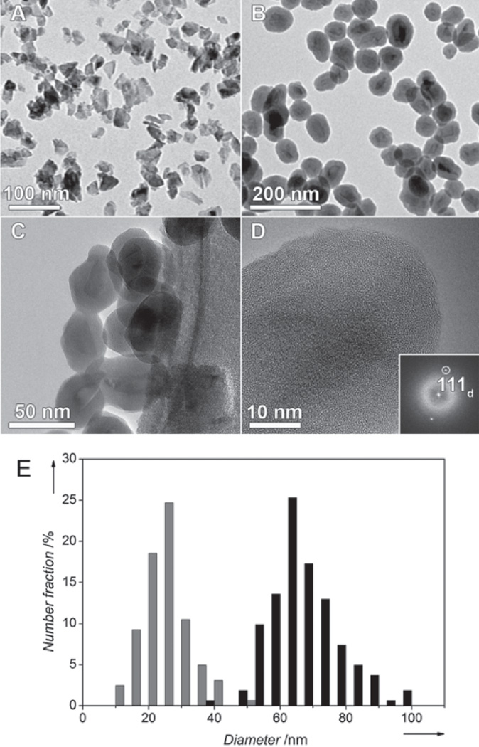
Figure 1.
BF-TEM images of non-coated ND1 (A) and aminosilica-coated ND3 (B) particles. Several coated ND3 particles are shown at higher magnification in (C). (D) High resolution TEM image showing the uniform silane surface coating. The crystallinity of the diamond core is evidenced by the presence of 111 diamond lattice planes, indexed in the Fourier Transform inset. (E) Histograms from image analysis of TEM micrographs of non-coated ND1 (gray) and aminosilica-coated ND3 (black) particles.

